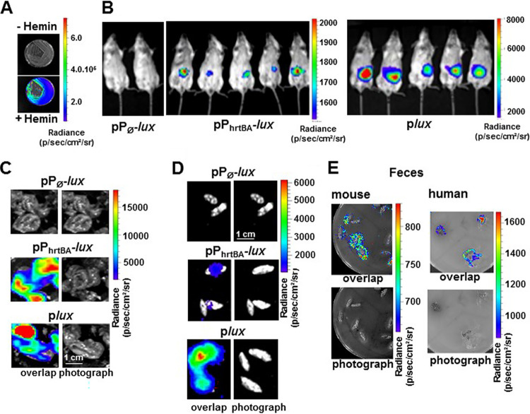
FIG 7.
HrtBAEf is induced in the gastrointestinal environment. (A) Heme-dependent light emission by the heme sensing WT(pPhrtBA-lux) strain. WT(pPhrtBA-lux) was plated on M17G agar plates in the presence (+) or absence (−) of 20 μM hemin. Plates were incubated at 37°C for 24 h, and luminescence was visualized using an IVIS 200 luminescence imaging system (acquisition time, 1 min; binning 8). Radiance is shown in photons per second per square centimeter per steradian. The results are representative of three independent experiments. (B) Heme sensing by E. faecalis over the course of intestinal transit. Female BALB/c mice were force fed with 108 CFU of WT(pPØ-lux) (control strain), WT(pPhrtBA-lux) (sensor strain), or WT(plux) (tracking strain). At 6 h postinoculation, anesthetized mice were imaged in the IVIS 200 system (acquisition time, 20 min; binning 16). The figure shows representative animals corresponding to a total of 15 animals for each condition in three independent experiments. (C) Ceca exhibit high heme sensing signal. Animals as described above for panel B were euthanized and immediately dissected. Isolated GITs were imaged as described above for panel B. Ceca that exhibited most of the luminescence are shown (acquisition time, 5 min; binning 8). Bar = 1 cm. (D) Visualization of heme sensing in feces collected from mice following ingestion of WT(pPØ-lux), WT(pPhrtBA-lux), or WT(plux). WT(pPhrtBA-lux) as described above for panel B were collected 6 to 9 h after gavage. Feces were imaged as described above for panel B (acquisition time, 20 min; binning 16). Bar = 1 cm. Results are representative of three independent experiments. (E) Human and mouse fecal samples activate heme sensing. Human feces from three healthy human laboratory volunteers and mouse feces from 6-month-old female BALB/c mice were deposited on M17G agar plates layered with soft agar containing WT OG1RF (pPhrtBA-lux). Plates were incubated at 37°C for 16 h and imaged in the IVIS 200 system (acquisition time, 10 min; binning 8). The figure shows representative results of a total of three independent experiments.

