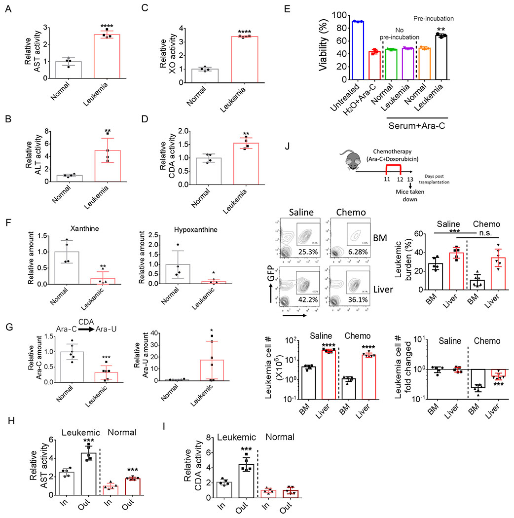
Figure 6.
Leukemic infiltration induces liver damage and creates a chemo-protective microenvironment.
A-D, AST (A), ALT (B), XO (C) and CDA (D) activities in sera from normal and leukemic mice (n=4).
E, Ara-C was preincubated with serum from normal or leukemic mice or H2O for 24 h at 37 ⁰C. One group of bulk leukemia cells were treated with preincubated Ara-C, the other group were treated with Ara-C without preincubation and supplemented with equal amount of either normal or leukemic serum as the first group. Viability of leukemia cells were examined 24 h after treatment. Error bars denote mean ± SD from triplicates.
F, Relative serum xanthine and hypoxanthine levels in normal and leukemic mice (n=4).
G, Ara-C was incubated with normal or leukemic serum for 24h at 37 ⁰C (final concentration of cytarabine: 100nM). Ara-C and Ara-U concentration were determined by mass spectrometry after incubation(n=6).
H-I, Plasma activity of AST (H) and CDA (I) in portal vein and hepatic vein from normal and leukemic mice (n=5).
J, Leukemic mice were treated with short-term chemotherapy (consisting of 2-day treatment of Ara-C (50 mg/kg, i.p.) and doxorubicin (1.5 mg/kg, i.p.). BM and liver leukemic burden, leukemic cell number (per whole liver or per femur + tibia) and leukemia cell number fold changed were determined (n=5).
Error bars denote mean ± SD. *p<0.05, **p<0.005, ***p<0.0005, ****p<0.00005.

