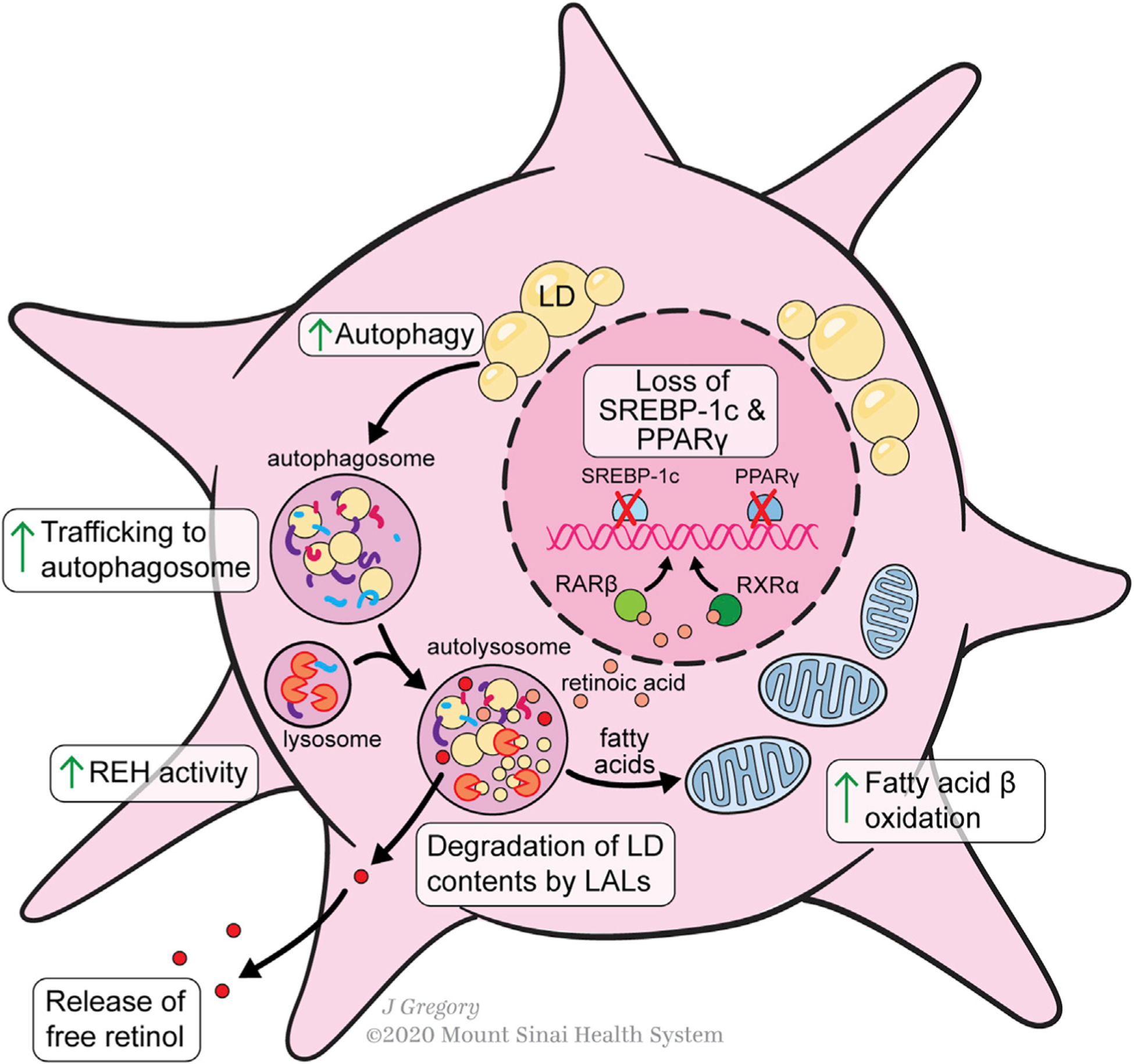
Figure 3. Lipid Metabolism during HSC Activation.

Lipid metabolism in activated HSCs is characterized by a loss of retinyl ester-containing cytoplasmic droplets. The transcription factors PPAR-γ and SREBP-1c, adipogenic master regulators, are markers of HSC quiescence that are downregulated during activation. Lipid droplets are trafficked to the autosome for degradation. Vitamin A de-esterification also occurs under the control of lysosomal acid lipase REH activity is also increased, liberating free retinol to the extracellular space. Lipid droplet contents from the autolysosome are further metabolized by the cell through increased fatty acid β-oxidation. Conversion of some intracellular retinol to retinoic acid promotes increased RARβ and RXRβ transcription.
