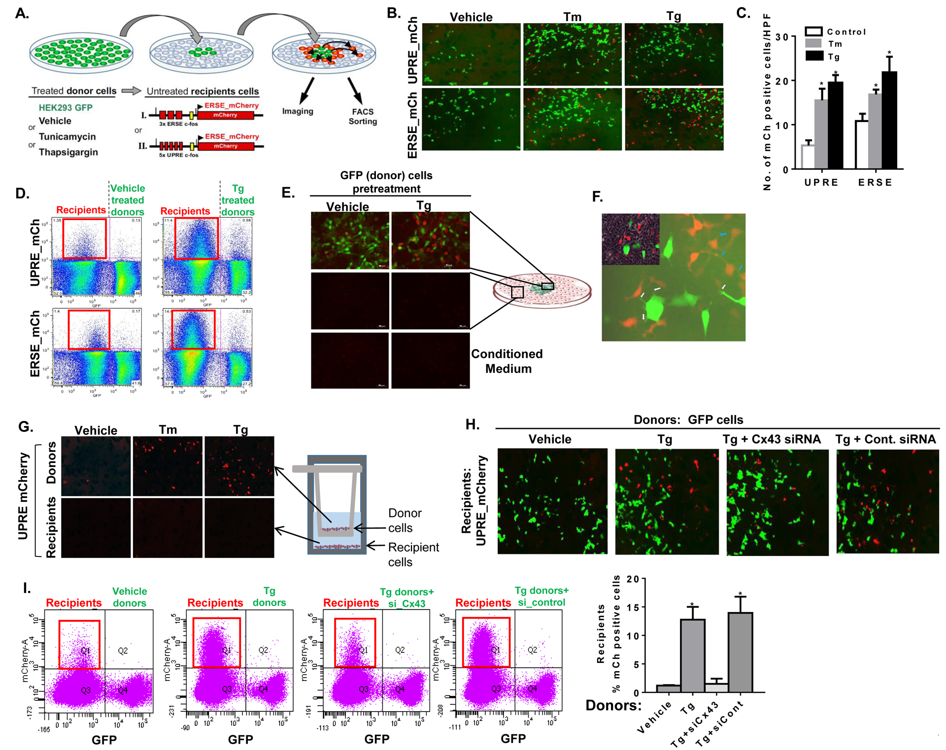
Figure 2: UPR is transmitted between cells in a Cx43-dependent manner.

(A) Illustration of the cellular models used to assess the transfer of ER stress signals from donor cells to ER stress naïve recipient cells. HEK293 cells were stably transfected with the red fluorescent protein mCherry driven by either the ER stress or the UPR response element (ERSE- and UPRE-mCherry). These cells express mCherry following exposure to ER stress and were used as ‘recipients’. GFP-expressing HEK293 cells were used as ‘donor’ cells and were pre-treated with either DMSO (Vehicle), tunicamycin (Tm) or thapsgargin (Tg). Following several washes, donor cells were transferred to a near confluent dish of ‘recipient’ cells and mCherry activation was assessed by fluorescent microscopy and by flow cytometry. (B) Donor GFP cells were pre-treated with either vehicle (DMSO), Tm (5 μg/mL) or Tg (200 nM) for 6 hours, followed by co-culture with either UPRE-mCherry (UPRE-mCh) or ERSE-mCherry (ERSE-mCh) recipient cells. Images of the mixed population of donor (GFP) and ER-stressed recipient (mCherry positive) cells were captured following 16 hours of co-culture. (C) Mean ± SD of mCherry (mCh) positive cells in 10 high power field (HPF) images of the mixed donor-recipient cell population. *p<0.05 as compared to vehicle. (D) Fluorescence-activated cell sorting (FACS) analysis of the mixed cell populations of GFP donor cells, pre-treated with either vehicle (left panels) or Tg (right panels) with UPRE-mCh (upper panels) or ERSE-mCh (lower panels) recipient cells following an overnight co-culture. The red square (left upper quadrant in each panel) highlights the extent of mCherry expression among recipient cells. (E) mCherry expression among UPRE-mCherry recipient cells was assessed by fluorescent microscopy within and around the GFP donor colony (upper panels) and remotely, at the periphery of the dish (middle panels). The effect of conditioned media from vehicle or Tg-treated donor cells on mCherry expression in recipient cells was also tested (lower panels). (G) A x400 magnification demonstrating thin cellular extensions of donor (GFP positive) to recipient (mCherry positive) cells (white arrows) and of recipient-to-recipient cells (blue arrows). Trans-well co-culture system was used to prevent cell-cell contact between donor and recipient cells, while incubated in the same medium. The ability of donor cells to transmit ER stress to the recipients was tested under these conditions. (H, I) mCherry expression in UPRE-mCherry recipient cells co-cultured overnight with vehicle (DMSO), or Tg-treated GFP donor cells. Donor cells were either transfected with Cx43 siRNA (si-Cx43) or with control siRNA (si-control). Fluorescent images (G) and FACS analyses (H) are presented. Q1 (framed in red) represents the extent of recipient cells expressing mCherry in each condition. Quantification of two independent experiments is presented, p<0.05.
