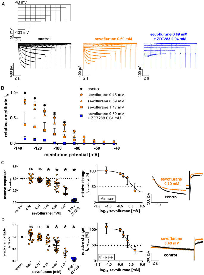
Figure 2.
(A) Representative current traces from thalamocortical relay neurons under control conditions with the corresponding voltage protocol, after application of 0.69 mM sevoflurane, and after applying both 0.69 mM sevoflurane and 0.04 mM ZD7288. (B) Beginning from a concentration of 0.45 mM, sevoflurane significantly reduced HCN-mediated Ih current amplitude over a broad range of membrane potentials. (C) The maximum Ih current amplitude (Ih maximal) is attained when the cell is hyperpolarized to −133 mV. The reduction of Ih maximal was dose-dependent. Corresponding current traces of Ih maximal under control conditions and in presence of 0.69 mM sevoflurane are depicted on the right. ns, not significant. *p < 0.05. (D) The reduction of Ih current amplitude in the presence of sevoflurane was also observable at membrane potentials closer to a neuron’s physiological state. Again, starting from a concentration of 0.45 mM, sevoflurane inhibited Ih current amplitude at −73 mV and this effect was concentration-dependent. Presented on the right, representative current traces of Ih at −73 mV under control conditions and in the presence of 0.69 mM.

