FIGURE 4.
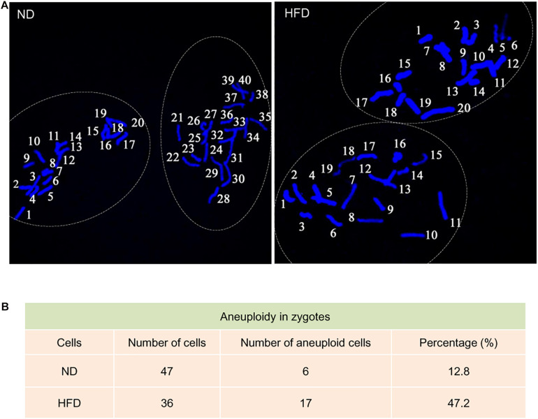
Increased aneuploidy in zygotes derived from HFD oocytes. (A) Chromosome spread of zygotes derived from ND and HFD oocytes (n = 47 ND zygotes from 5 mice and n = 36 HFD zygotes from 5 mice). Blue, chromosomes stained with DAPI. Representative confocal images show the euploidy in ND zygotes and aneuploidy in HFD zygotes. (B) Summary of the frequency of aneuploidy in ND and HFD zygotes.

