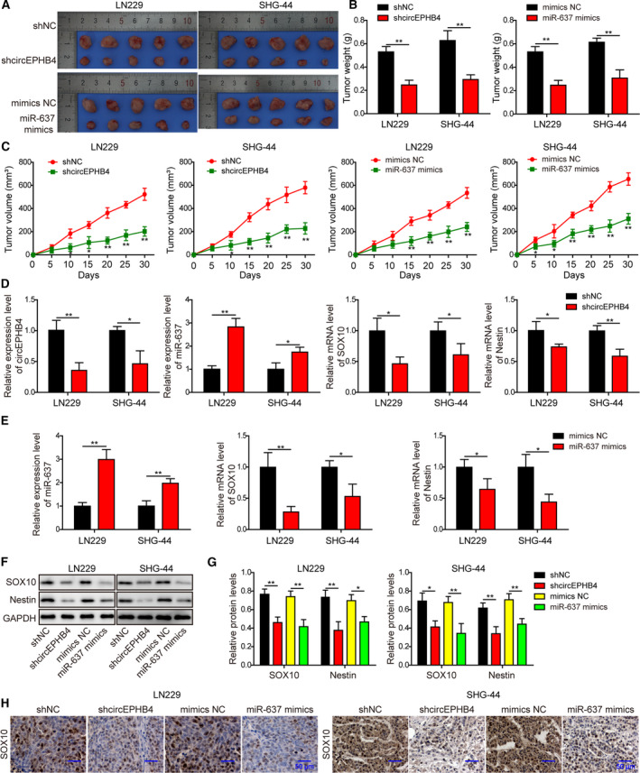
Fig. 14.

Knocking down circEPHB4 or up‐regulating miR‐637 inhibited the in vivo xenograft growth of glioma cells. (A) Images of xenograft tumors from indicated groups. (B) Weights of all xenograft tumors were measured and compared between indicated groups. (C) The growth curve of xenograft tumors is presented as changes in tumor volumes from indicated groups. (D) The expression levels of circEPHB4, miR‐637, SOX10 and Nestin were examined by qRT‐PCR in xenograft tumors derived from indicated cells. (E) The expression levels of miR‐637, SOX10 and Nestin were examined by qRT‐PCR in xenograft tumors derived from indicated cells. (F,G) The expression levels of SOX10 and Nestin were examined by Western blotting in xenograft tumors derived from indicated cells. The representative images are shown in (F) and quantified as the ratio to GAPDH (G). (H) Histological analysis on SOX10 from indicated xenograft tumors. Scale bar: 50 µm. LN229 or SHG‐44 cells expressing shcircEPHB4 or miR‐637 mimics were subcutaneously injected into nude mice (n = 5/group). After 30 days, all mice were euthanized. Data are presented as mean ± SD from three independent experiments. Comparison between two groups was performed using Student’s t‐test and between three or more groups using one‐way ANOVA followed by Tukey’s post hoc test. *P < 0.05, **P < 0.01.
