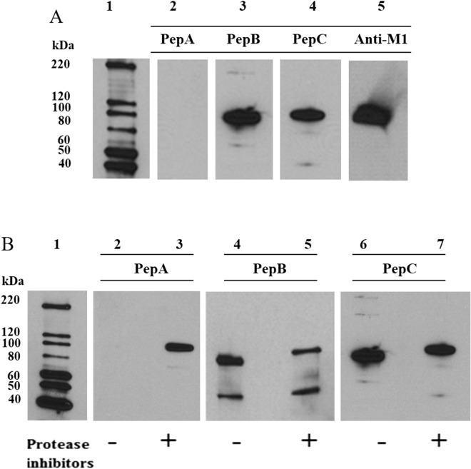
Figure 3.
Immunoblots with peptide antibodies. (A) Recombinant PfM1AAP was probed with antibodies prepared against 14-mer peptides derived from the N-terminal extension (PepA), spanning domain 1 (PepB) and domain 4 (PepC) (see Supplementary Fig. 1). Anti-PfM1AAP was used as a positive control. Anti-PepA does not react with rPfM1AAP, as this lacks the N-terminal extention, whereas both anti-PepB and anti-PepC do. Lane 1 was loaded with chemilluminescent molecular marker. Lanes 2–5 were loaded with 0.25 µg of recombinant M1. (B) Cytosolic fractions of malaria parasites prepared without (−) and with (+) a protease inhibitor cocktail were probed with anti-PepA, anti-PepB and anti-PepC antibodies. Extracts were separated on 4–15% SDS-PAGE gels and transferred to nitrocellulose membrane. Lanes 2 and 3 were probed with anti-PepA, lanes 4 and 5 with anti-PepB and lanes 6 and 7 with anti-PepC antibodies. The chemiluminescent molecular marker is shown in lane 1.

