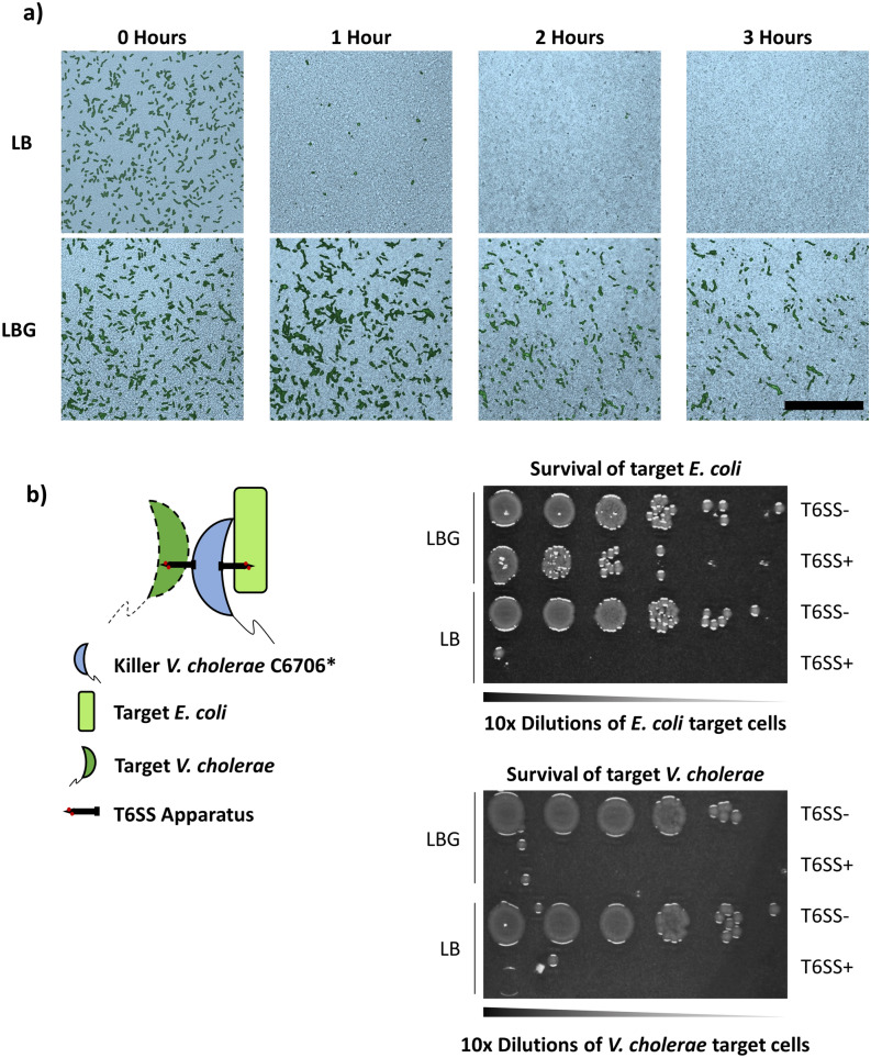
Figure 3.
Individual E. coli MG1655 cells are protected against V. cholerae T6SS attacks but do not impair T6SS killing. (a) Fluorescently labeled green E. coli MG1655 cells were densely co-cultured with unlabeled V. cholerae C6706* on LB (top panels) or LBG (bottom panels) and the same frame was imaged for three hours using confocal microscopy. The pseudocolor images depict the progression of T6SS killing at different time points, showing brightfield microscopy images (bright blue) of the dense biofilm overlaid with the fluorescence signal of E. coli cells projected on a plane parallel to the agar substrate. Scale bar is 50 µm. (b) Killer V. cholerae C6706* or C6706* T6SS- was co-cultured with both E. coli MG1655 and susceptible V. cholerae target cells, and then diluted and plated as described in the “Methods” section.

