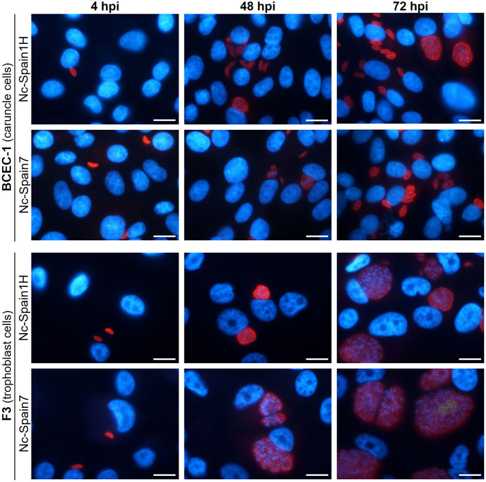
Figure 4.
Tracing the lytic cycle of high- (Nc-Spain7) and low-virulence (Nc-Spain1H) isolates of N. caninum in BCEC-1 and F3 cells at 4, 48, and 72 h post-infection (hpi) through immunofluorescence assays. Host nuclei were stained in blue, while N. caninum tachyzoites were stained in red. Note the obvious differences in the parasitophorous vacuole size between BCEC-1 and F3 cells infected with any of the isolates, which is due to an early egression of the parasites in the first. While no differences were observed in parasitophorous vacuole size between BCEC-1 cells infected with Nc-Spain1H or Nc-Spain7 tachyzoites, remarkable differences were evidenced in F3 cells, with bigger vacuoles in Nc-Spain7 infected cells compared to those infected with Nc-Spain1H. Note the beginning of egress in F3 cells infected with the Nc-Spain7 isolate at 72 hpi (stained in green). Scale-bars: 10 μm.

