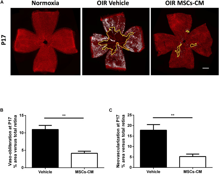
FIGURE 2.
Conditioned media (CM) of hypoxic MSC (MSCs-CM) promoted vascular growth in OIR retinas. (A) Representative photomicrographs from isolectin B4-stained retinal flatmounts from normoxic or OIR mice at P17 intravitreally treated with vehicle or MSCs-CM. Scale bar 500 μm. Retinas intravitreally injected with MSCs-CM demonstrate decreased VO (B) and NV (C) areas highlighted in yellow and white, respectively. Quantification of VO and NV is represented in the graphs (**p < 0.01 vs vehicle, values are mean ± SEM, n = 6–10 retinas).

