FIGURE 6.
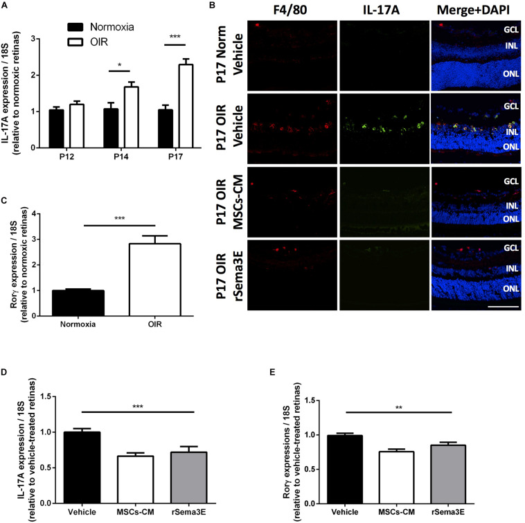
MSCs-CM downregulated IL-17A levels in retinal myeloid cells. (A) qPCR analysis of IL-17A in whole retina from mice pups at different time points of OIR demonstrating increased levels of IL-17A mRNA by P14 and P17 (*p < 0.05, ***p < 0.001 vs normoxia, values are mean ± SEM, n = 4–5, pool of 2 retinas per n). (B) Representative cryosections of normoxic and OIR retinas treated with Vehicle, MSCs-CM or rSema3E demonstrating co-localization of IL-17A with myeloid cell F4/80 marker. Nuclei were counterstained with DAPI (blue). NFL, nerve fiber layer; GCL, ganglion cell layer; INL, inner nuclear layer; ONL, outer nuclear layer. Scale bar 100 μm. (C) Real-time quantification (qPCR analysis) of P17 OIR retinas demonstrated increased expression of the nuclear receptor RORγ which regulates IL-17A transcription (***p < 0.001, values are mean ± SEM, n = 4–5, pool of 2 retinas per n). (D) Intravitreal injection of OIR retinas with MSCs-CM and rSema3E demonstrated lower levels of IL-17A at P17 versus vehicle-injected OIR retinas (***p < 0.001, n = 3–4, pool of 2 retinas per n). (E) Intravitreal injection of MSCs-CM and rSema3E exhibited significant decreased RORγ expression in OIR retinas via qPCR analysis at P17 in comparison to vehicle-treated counterpart (**p < 0.01, values are mean ± SEM, n = 4–5, pool of 2 retinas per n).

