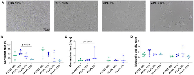
Figure 5.
(A) Representative phase-contrast photomicrographs of equine mesenchymal stromal cells (MSC) from one donor at day 5 after seeding for population doubling assays in media supplemented with fetal bovine serum (FBS) or equine platelet lysate (ePL). (B–D) Confluent area at day 5, measured using Fiji ImageJ (B), generation times calculated from cell doubling numbers (C), and metabolic activity as determined by MTS tetrazolium-based cell proliferation assay (D) for MSC in passage 2 (P2) cultured in the different media; Friedman and Wilcoxon tests for group comparisons were performed (p-values are indicated); the plots display the individual values, median, and interquartile ranges. Data were obtained from MSC from n = 4 donors. Note that missing generation time data are due to the lack of proliferation in these samples (1 out of 4 in the 5% ePL group, 4 out of 4 in the 2.5% ePL group).

