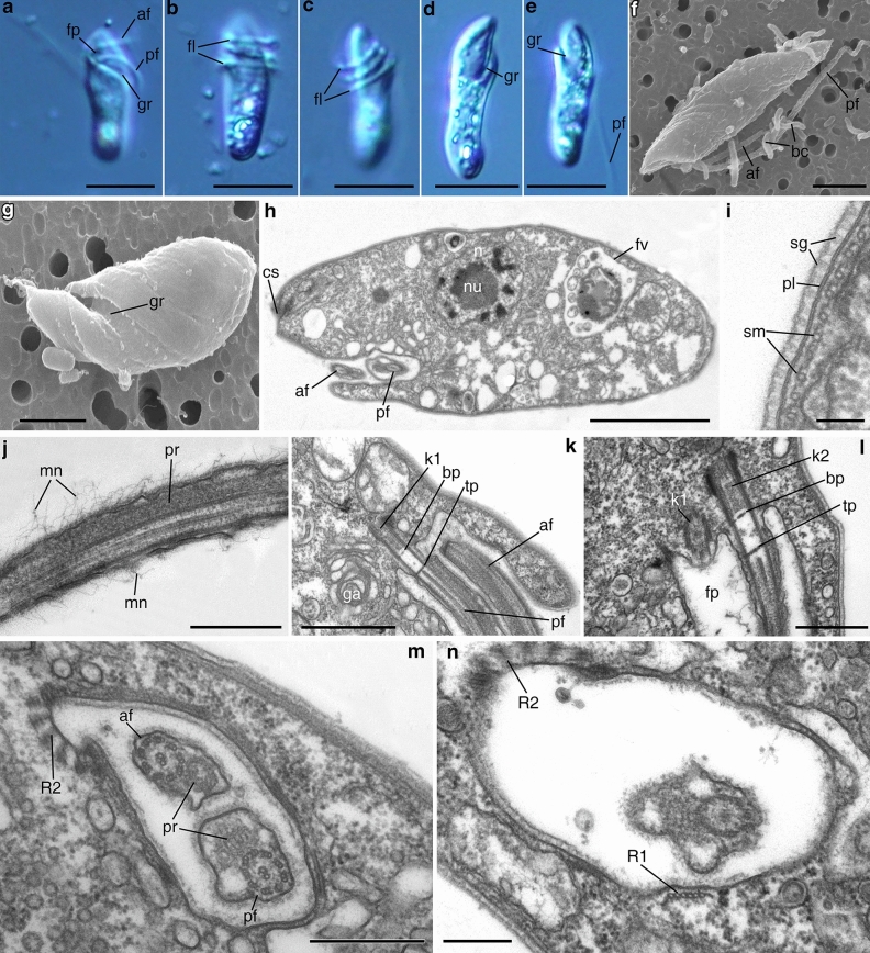
Figure 2.
General view, cell coverings, and flagellar pocket of Papus ankaliazontas, clone PhM-4. (a–e) living cells, LM, DIC; (f, g) general cell view, SEM (g flagella are not visible); (h) longitudinal cell section; (i) coverings structure; (j) anterior flagellum; (k, l) arising of the flagella from flagellar pocket; (m, n) cross sections of the flagellar pocket. af anterior flagellum, bc bacterium, bp basal plate, cs cytostome, fl flagella, fp flagellar pocket, fv food vacuole, ga Golgi apparatus, gr groove, k1 kinetosome (basal body) of the posterior flagellum; k2 kinetosome (basal body) of the anterior flagellum, mn mastigonemes, n nucleus, nu nucleolus, pf posterior flagellum, pl plasmalemma, pr paraflagellar rod, R1 mucrotubular root R1, R2 mucrotubular root R2, sg structured glycocalyx, sm subpellicular microtubules, tp transversal plate. Scales: (a–e) 10, (f) 3, (g) 2, (h) 3, (i) 0.1, (j) 0.5, (k) 1, (l–m) 0.5, (n) 2.5 μm.

