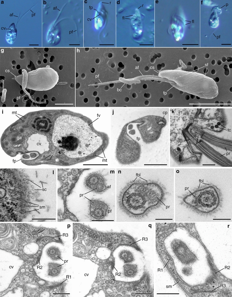
Figure 4.
General view, cell coverings and arrangement of flagellar pocket of Apiculatamorpha spiralis (a–j,l–r—clone PhF-6, k—clone PhF-5). (a–f) living cell, LM, DIC; (g,h) general cell view, SEM; (i) longitudinal section of the cell, (j) transverse section of the anterior cell end; (k) arising of the flagella from flagellar pocket; (l) scales on the cell surface; (m–o) cross sections of the flagella (n—posterior flagellum, o—anterior flagellum); (p–r) microtubular roots and flagellar pocket. af anterior flagellum, bc bacterium, bp basal plate, cp cytopharynx, cs cytostome, cv contractile vacuole, db dorsal band of the microtubules, fbl fiber layer, fl flagella, fp flagellar pocket, fv food vacuole, mt mitochondrion, n nucleus, nu nucleolus, pf posterior flagellum, p prey, pr paraflagellar rod, r rostrum, R1 microtubular root R1, R2 microtubular root R2, R3 microtubular root R3, sf spiral furrow, sc scales, sm subpellicular microtubules, tc trichocyst, tp transversal plate. Scales: (a–f) 5, (g) 2, (h) 5, (i,j) 2, (k) 0.5, (l) 0.2, (m) 0.5, (n,o) 0.2, (p–r) 0.5 μm.

