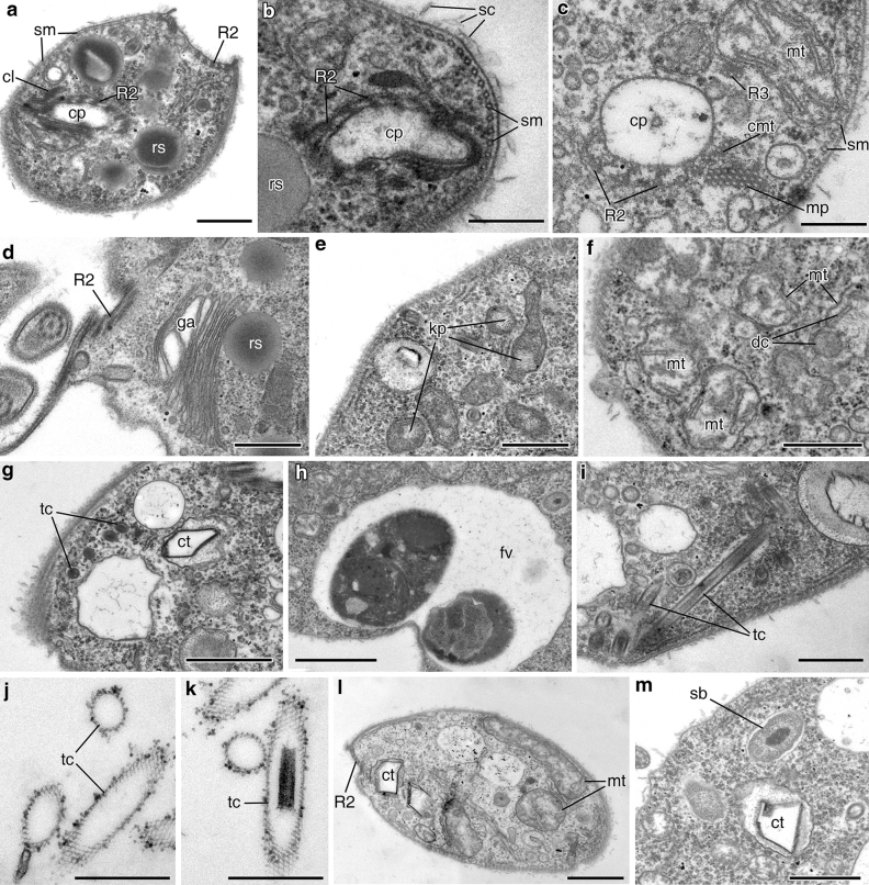
Figure 5.
Arrangement of cytopharynx and cell structures of Apiculatamorpha spiralis, clone PhF-6, including trichocysts. (a) section of the anterior cell end with cytopharynx; (b, c) cytopharynx and accociated structures; (d) Golgi apparatus; (e, f) mitochondria and kinetoplasts; (g) trichocysts and crystalloid structure; (h) food vacuole; (i) longitudinal section of the trichocysts; (j, k) empty envelopes of the trichocysts after discharging; (l) crystalloid structure and mitochondria; (m) symbiotic bacterium and crystalloid structure. cl clamp, cmt cytopharynx associated additional microtubules, ct crystalloid structure, cp cytopharynx, dc discoid cristae, fv food vacuole, ga Golgi apparatus, kp kinetoplast, mt mitochondrion, mp microtubular prism, R2 microtubular root R2, R3 microtubular root R3, rs reserve substance, sm subpellicular microtubules, sb symbiotic bacterium, sc scales, tc trichocyst. Scales: (a) 0.5, (b,c) 0.2, (d–f) 0.5, (g,h) 1, (i–k) 0.5, (l) 1, (m) 0.5 μm.

