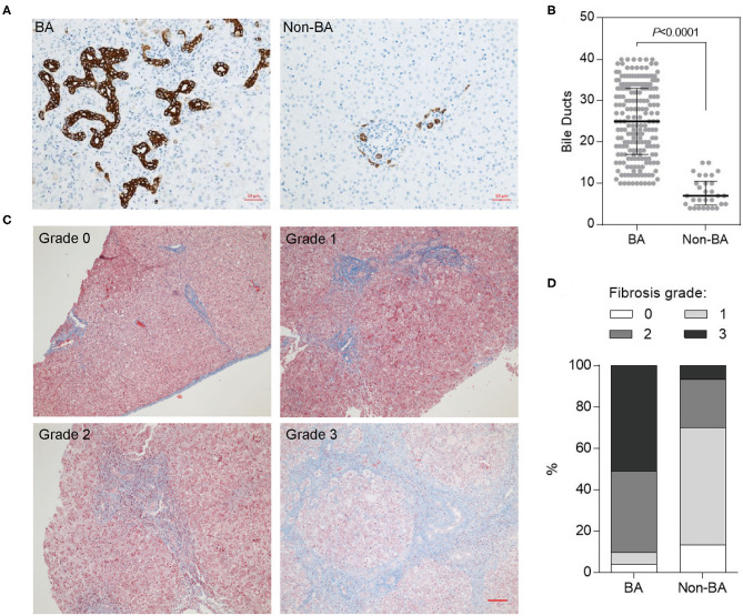
Figure 1.
Bile duct hyperplasia and fibrosis in BA livers of Chinese patients. (A) Hyperplastic and deformed bile ducts in BA livers indicated by CK19 IHC staining. Scale bar: 50 μm. (B) Quantification of (A) (numbers of bile duct per field). (C) Fibrosis in BA livers indicated by Masson's trichrome staining. Scale bar: 100 μm. (D) Fibrosis grades in BA and Non-BA livers were summarized.

