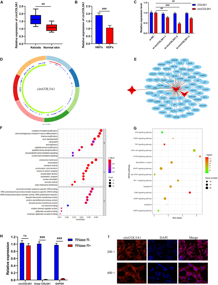
FIGURE 1.
Bioinformatics analysis and characterization of circCOL5A1. (A) The relative RNA levels of circCOL5A1 were evaluated by qRT-PCR between keloid tissues and normal skin. (B) The relative RNA levels of circCOL5A1 were evaluated by qRT-PCR between HKFs and HDFs. (C) The silent efficiency of circCOL5A1 was evaluated by qRT-PCR in HKFs transfected with si-NC or siRNAs, respectively. (D) The structure and binding sites of circCOL5A1. The red sites represented the microRNA response element. The blue sites represented RNA binding protein. The green sites represented an open reading frame. (E) Construction of circRNA-miRNA-mRNA network. GO (F) and KEGG (G) analysis of circCOL5A1 target genes. (H) The relative abundance of circCOL5A1 or linear COL5A1 in HKFs were detected by qRT-PCR after treatment with or without RNase R. (I) FISH assays were performed to observe the cellular location of circCOL5A1 (red) in HKFs (magnification, 200× and magnification, 400×). ##p < 0.01 and ###p < 0.001.

