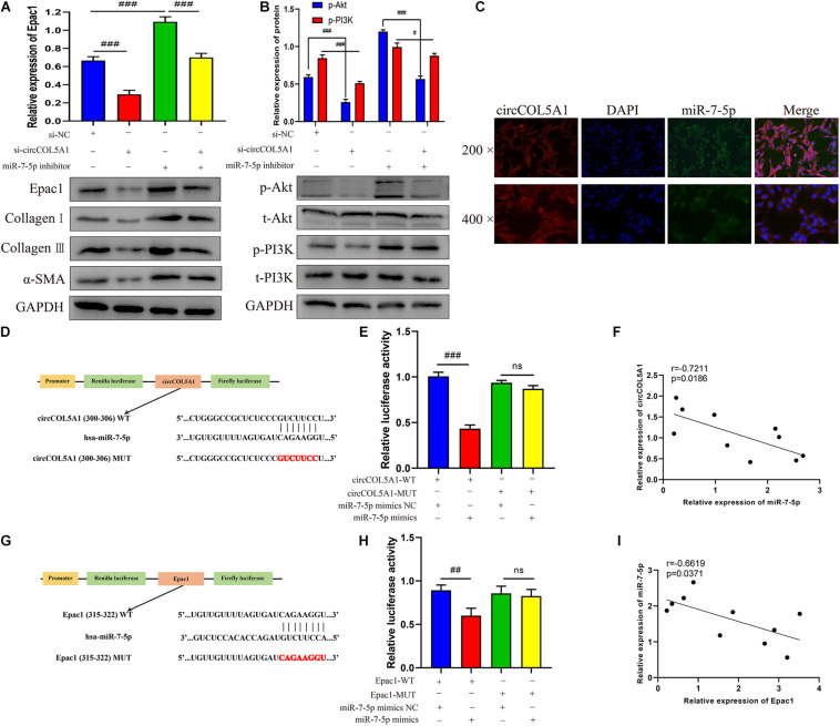
FIGURE 4.
circCOL5A1 served as a miRNA sponge of miR-7-5p to regulate Epac1 expression. (A,B) The protein levels of collagen I, collagen III, α-SMA, and the protein phosphorylation levels of Akt and PI3K in HKFs transfected with si-NC, si-circ, or si-circ + inhibitor were determined using western blot, respectively. (C) FISH assays were performed to observe the cellular location of circCOL5A1 (red) and miR-7-5p (green) in HKFs (magnification, 200× and magnification, 400×). (D) Schematic diagram of circCOL5A1-WT and circCOL5A1-MUT luciferase reporter vectors. (E) The relative luciferase activities were evaluated in HKFs after co-transfection with circCOL5A1-WT or circCOL5A1-MUT and mimics or NC, respectively. (F) Pearson correlation analysis was performed to evaluate the correlation between circCOL5A1 and miR-7-5p in keloid tissues. (G) Schematic diagram of miR-7-5p-WT and miR-7-5p-MUT luciferase reporter vectors. (H) The luciferase activity of reporter that carried WT rather than Mut 3′-UTR of Epac1 was markedly suppressed by miR-7-5p mimics. (I) Pearson correlation analysis was performed to evaluate the correlation between miR-7-5p and Epac1 in keloid tissues. Data was shown as mean ± SD. ns indicated no significance, #P < 0.05, ##P < 0.01, ###P < 0.001.

