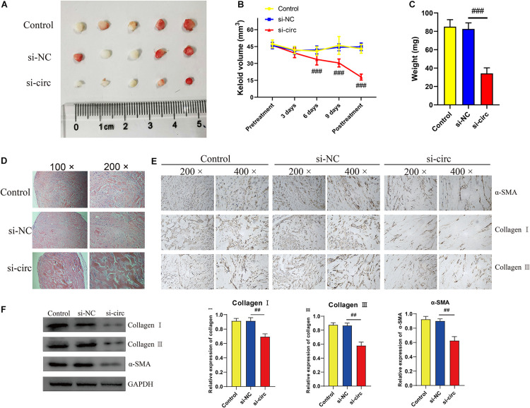
FIGURE 6.
Downregulation of circCOL5A1 suppressed the growth of keloids and ECM deposition in vivo. (A) Images of subcutaneous keloid grafts in circCOL5A1 low expression group and control group. (B) The relative volume of keloid grafts was analyzed. (C) The weight of the keloid grafts was evaluated. (D) Representative images of HE staining of keloid nodules in different intervention groups (magnification, 100× and magnification, 200×). (E) The relative expression level of collagen I, collagen III, and α-SMA was observed in keloid grafts by IHC (magnification, 200× and magnification, 400×). (F) The protein levels of collagen I, collagen III, and α-SMA were evaluated in subcutaneous keloid grafts by western blot analysis. Data was shown as mean ± SD. ns indicated no significance, ##P < 0.01, ###P < 0.001, vs. si-NC.

