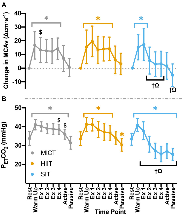
FIGURE 2.
(A) MCAv was determined continuously by transcranial doppler ultrasound (TCD) and presented as change in MCAv from rest in each protocol for each participant. (B) PETCO2 was determined through breath-by-breath respiratory gas exchange and ventilation. Data were averaged over 30 s of rest (rest); 5 min warm up (warm up); 3 min active recovery (active); and 15 min seated passive recovery (passive) in all protocols. Exercising averages (Ex 1–4) were taken every 7 min during 30 min of moderate intensity (65% VO2peak) steady state exercise (MICT); in the final minute of the four 4 min high intensity (85% HRmax) interval bouts (HIIT) and across the duration of four 30 s supramaximal (200% Wmax) sprint intervals (SIT). Data are presented as mean ± SD (n = 24; except SIT–Passive: n = 23) and were analyzed by linear mixed models. Significant differences (analyzed by linear mixed model, p < 0.05) between resting values and subsequent time points are denoted by *. Significant differences between protocols are denoted by $ between MICT and HIIT; † between MICT and SIT; and Ω between HIIT and SIT.

