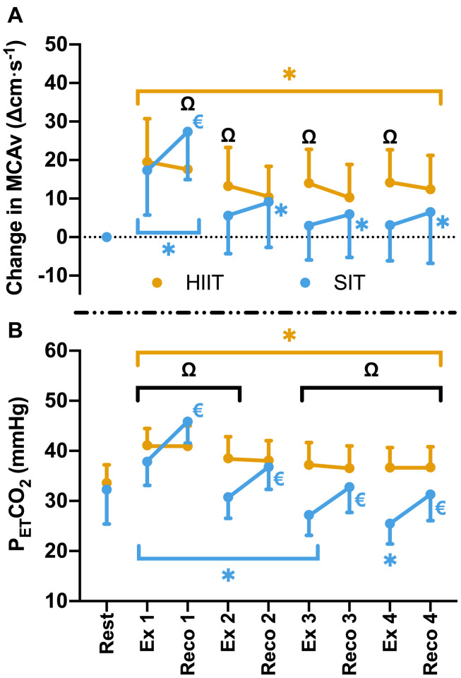
FIGURE 3.
(A) MCAv was determined continuously by transcranial doppler ultrasound (TCD) and presented as change in MCAv from rest in each protocol for each participant. (B) PETCO2 was determined by measurement of breath-by-breath respiratory gas exchange and ventilation. Data were averaged for 1 min during seated rest (Rest); over 30 s during the final minute of four 4 min high intensity (85% HRmax) interval bouts (HIIT; Ex 1 – 4); across the duration of four 30 s supramaximal (200% Wmax) sprint intervals (SIT; Ex 1 – 4), and over a 30 s period, 15 s into the recovery following each bout in both interval protocols (Reco 1 – 4). Data are presented as mean ± SD (n = 24). Significance (analyzed by linear mixed model, p < 0.05) between time points is denoted by * for differences from resting values; € for differences between interval (ex) and recovery (reco). Significant differences between protocols are denoted by Ω.

