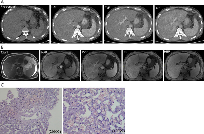
Figure 3.
Representative CT, MRI, and histological imaging. (A) Representative CT image. (B) Representative MRI image; 49-year-old man with Gynura segetum-induced HSOS received contrast-enhanced CT and a gadoxetic acid-enhanced MRI scan. Heterogeneous hypoattenuation/hypointensity and patchy enhancement are shown. (C) Representative histological results; 36-year-old man with Gynura segetum-induced HSOS received liver biopsy, with the results showing massive sinusoidal dilatation and sinusoidal congestion accompanied by the extravasation of erythrocytes into the space of Disse. HAP, hepatic arterial phase; PVP, porta-venous phase; EP, equilibrium phase; HBP, hepatobiliary phase; HSOS, hepatic sinusoidal obstruction syndrome.

