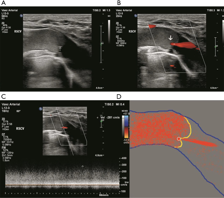
Figure 1.
Duplex ultrasonography showed the location of the membrane occlusion of the SCV. (A) Duplex ultrasonography demonstrated a fornix-shaped membrane (white arrow) located at the proximal end of the right SCV. The blood flow was slow distal to the membrane. (B) Color Doppler ultrasound showed the blood flow being forced out from the small orifice (white arrow) of the membrane. (C) The maximal velocity of blood flow at the orifice was up to 281 cm/s. (D) Sketch of the SVC, fornix-shaped membrane and orifice.

