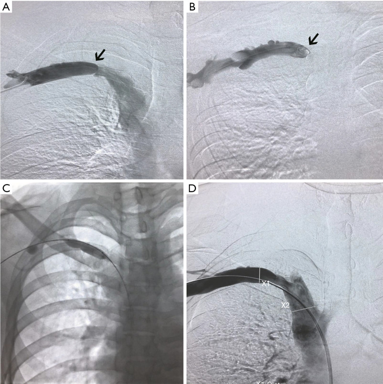
Figure 3.
Venography of the SCV indicated the blood flow before and after the dilation balloon. (A,B) Venography indicated a fornix-shaped membrane (black arrow) at the proximal segment of the SCV without signs of thrombosis. The blood flow was almost completely obstructed by the membrane. There were no obvious collaterals around the occlusion. (C) The membrane was gradually dilated using an 8–40 mm balloon and a 12–40 mm balloon. (D) After dilation, the blood flow in the SCV was recovered with residual stenosis of less than 30%. SCV, subclavian vein.

