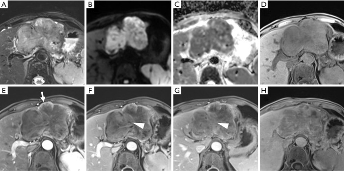
Figure 7.
A histopathologically confirmed poorly differentiated hepatocellular carcinoma in a 71-year-old male patient with chronic hepatitis B. A mass in left lobe of the liver shows mild-to-moderate T2 hyperintensity (A), restricted diffusion (B,C), hypointensity on pre-contrast image (D), rim arterial phase hyperenhancement (E) (arrow), delayed central enhancement (F,G) (triangle), and hepatobiliary phase hypointensity (H). These imaging features would lead to incorrect designation as LR-M, suggesting intrahepatic cholangiocarcinoma.

