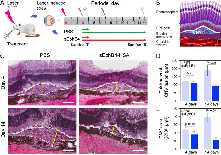
Figure 3.
Effects of sEphB4-HSA injections on rat CNV (choroidal neovascularization) formation induced by laser. (A) Schematic diagram of the timeline of the laser application in rats, treatments, and sacrifice. (B) Morphology scheme of laser-induced CNV in rat. (C) A representative light microscopic images of CNV lesion were obtained at days 4 or 14 after laser application. Hematoxylin & eosin stain (H&E) (C), yellow line indicates the maximum thicknesses of CNV and CNV areas are outlined with white dot line. Scale bar: 100 µm. statistical analysis of thickness of rat CNV lesion (D) and CNV area in (E).

