
Figure 1.
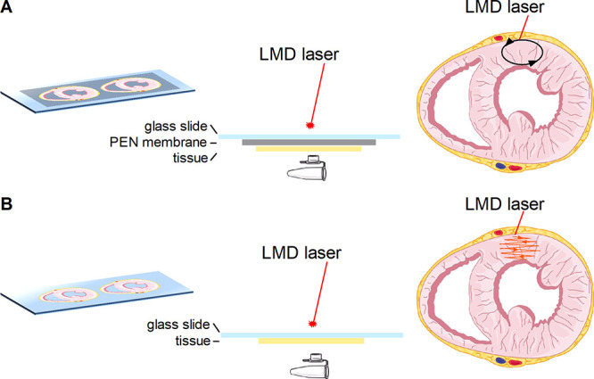
Schematic representation of the dissection method used for LMD. The selected region is dissected from a PEN membrane slide [(A) black oval] or ablated from a conductive, nonmembrane slide [(B) orange pattern]. Graphical elements were adapted from Servier Medical Art.