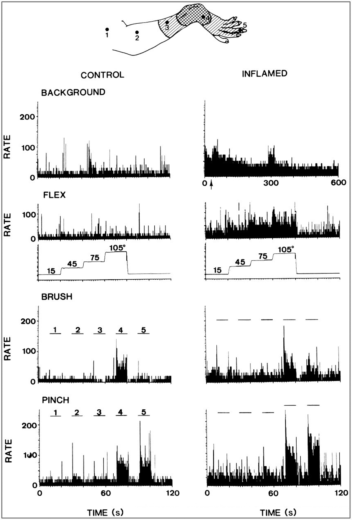
FIG. 4.

Increased responses of a primate spinothalamic tract cell after acute arthritis was induced by injection of kaolin and carrageenan into the knee joint. Top: Receptive field of the neuron on the ankle and foot before (doubly hatched area) and after(hatched area) the development of arthritis. Left columns: Peristimulus histograms show the background activity of the neuron and its responses to flexion of the knee, to brushing the skin at the points labeled 1–5 in the drawing, and to pinching the skin at the same points. Right columns: Histograms show the enhanced background activity and response after the development of arthritis. The increased background activity would presumably result in pain in an unanesthetized animal, and the increased response to knee flexion would be an indication of primary hyperalgesia. The increased responses to stimulation of the foot would presumably represent secondary mechanical allodynia and hyperalgesia. (From Dougherty et al., 1992b.)
