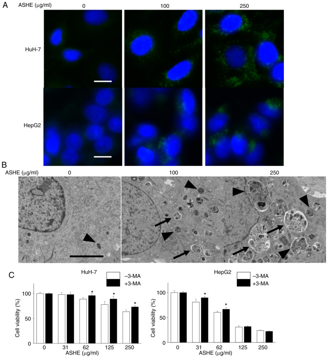
Figure 4.
Autophagic vesicle quantity is increased in ASHE-treated liver cancer cells. (A) Fluorescence micrographs of HuH-7 and HepG2 cells treated with ASHE in the absence of FBS for 72 h. Following treatment, cells were incubated with DAPGreen (50 nM) for 30 min. Magnification, ×400. Scale bar, 10 µm. (B) Representative transmission electron micrographs of HuH-7 cells treated with ASHE in the absence of FBS. Arrows indicate autophagosomes, and arrowheads indicate autolysosomes. Magnification, ×5,000. Scale bar, 5 µm. (C) ASHE-treated HuH-7 and HepG2 cells were co-treated with 3-MA (0.1 or 0.3 mM) for 72 h, followed by measurement of cell viability. Data are presented as the mean ± standard deviation of three independent experiments. *P<0.05 vs. ASHE-treated cells in the absence of 3-MA. ASHE, Acanthopanax senticosus Harms root extract; 3-MA, 3-methyladenine.

