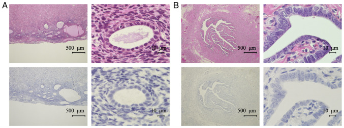
Figure 5.
IHC analysis of TFPI-2 in non-neoplastic tissues. Representative images of H&E staining (top) and IHC for TFPI-2 (bottom) in non-neoplastic tissues. (A) Endometrium of TFPI-2-negative case. (B) Fallopian tube epithelium. IHC, immunohistochemistry; TFPI-2, tissue factor pathway inhibitor-2; H&E, hematoxylin and eosin.

