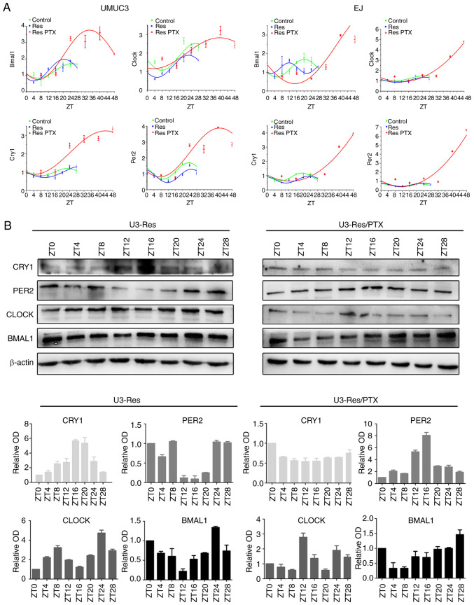
Figure 2.
Res cells enter quiescence with prolonged circadian period. After cells (UMUC3 and EJ, Res and Res + PTX) were synchronized with 0.1 µM dexamethasone for 2 h, circadian genes were detected at different time points. (A) mRNA expression levels of circadian clock genes (BMAL1, CLOCK, PER2 and CRY1) and corresponding fitted cosinor curves. (B) Time course of protein (BMAL1, CLOCK, PER2 and CRY1) expression levels in the Res and Res + PTX (80 nM) cells. β-Actin was used as loading control. Data are presented as the OD fold difference related to the control from three duplicate experiments. Res, cisplatin-resistant cells; PTX, paclitaxel; OD, optical density; ZT, circadian time; CRY1, Cryptochrome 1; PER2, period 2; CLOCK, circadian locomotor output cycles kaput; BMAL1, brain and muscle Arnt-like protein 1.

