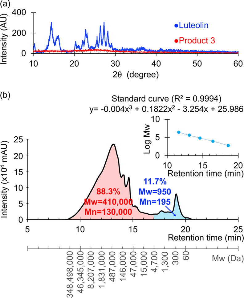
Figure 3.

(a) X-ray diffraction (XRD) patterns of luteolin and polyluteolin nanoparticles (Product 3) and (b) gel filtration chromatography (GFC) chromatogram (10 mg/mL, N,N-dimethylformamide (DMF); PDA detector at 254 nm) of polyluteolin (Product 3). Polystyrene (Mp = 580, 9820, 67600, 466300, and 3152000 Da) standard curve is shown in the upper left.
