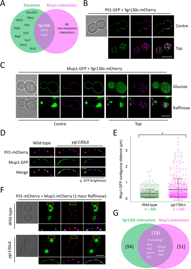
Fig. 6.
Ygr130c is required for efficient eisosomal cargo retention. (A) Venn diagram showing overlap between known eisosome proteins (green) and Mup1 physical interactors (magenta). (B) Cells co-expressing Pil1–GFP (green) and Ygr130c–mCherry (magenta) were imaged at centre and top focus planes using confocal microscopy. Merge images are shown on the right. (C) Cells expressing Ygr130c–mCherry (magenta) were transformed with a Mup1–GFP (green) plasmid and imaged in glucose or raffinose media by Airyscan confocal microscopy. Merge images are shown on the right for each condition. Arrows indicate spots of colocalisation. (D) Colocalisation of Mup1–GFP- and Pil1–mCherry-marked eisosomes in wild-type and ygr130cΔ mutants was recorded by Airyscan confocal imaging. A panel of reduced GFP brightness is included to better show GFP distribution (right). (E) Contiguous plasma membrane GFP signal (in µm) was measured in wild-type (green) and ygr130cΔ cells (magenta). Mean±s.d. (F) Wild-type and ygr130cΔ cells co-expressing Pil1–mCherry (magenta) and Mup1–GFP (green) following 1 h of raffinose exchange were compared using Airyscan microscopy. Boxes indicate regions shown in enlarged images below. (G) Venn diagram between physical interactors of Ygr130c (green) and Mup1 (magenta); 18 proteins are present in both sets. *P<0.05 (Student's t-test). Scale bars: 5 μm (white), 1 μm (yellow).

