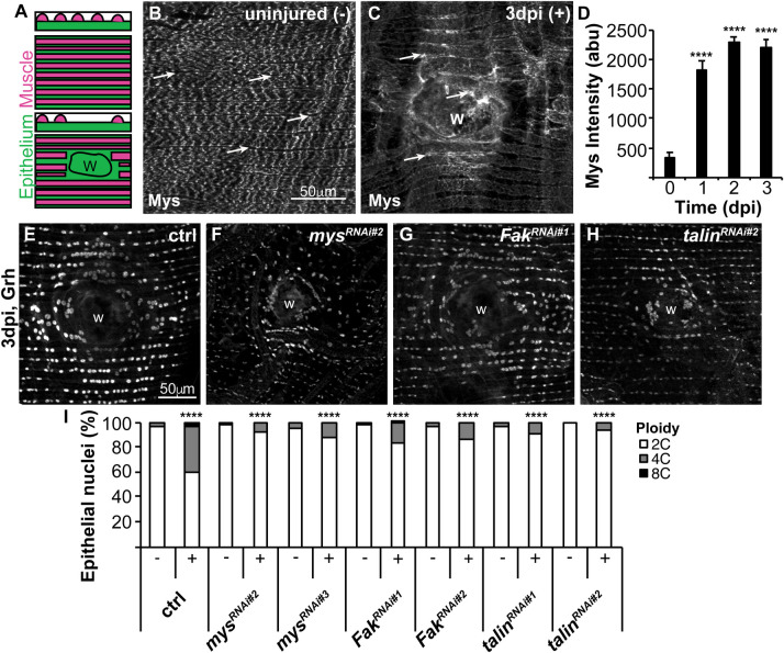
Fig. 1.
Focal adhesion genes are induced and required for endoreplication. (A) Illustration of the adult Drosophila abdominal organization of the lateral muscle fibers (red), overlaying the epithelium (green) in the transverse, z-view (top) and flattened, x-y view (bottom). Epithelial gene expression can be observed and measured in the gaps between overlaying muscle fibers. After injury the epithelium, but not the muscle fibers are repaired over the wound scar (outlined, w). (B) Representative immunofluorescent images of mys staining in the (B) uninjured (−) and (C) 3 dpi (+) adult fly abdomen. Epithelial mys expression is marked by arrows. Wound site, w. (D) Time course of mys expression quantified in the epithelium at 0 dpi (n=5), 1 dpi (n=12), 2 dpi (n=13), and 3 dpi (n=12). Error bars represent mean±s.e. and data were analyzed by two-tailed unpaired t-test. (E–H) Representative immunofluorescent images of control, mysRNAi, FakRNAi, and talinRNAi at 3 dpi stained with epithelial nuclear marker (Grh). (I) Quantification of epithelial ploidy in the control (−, n=15 and +, n=12), mysRNAi#2 (−, n=12 and +, n=10), mysRNAi#3 (−, n=11 and +, n=9), FakRNAi#1 (−, n=12 and +, n=8), FakRNAi#2 (−, n=7 and +, n=7), talinRNAi#1 (−, n=5 and +, n=4), and talinRNAi#2 (−, n=4 and +, n=3). Data were analyzed by two-way ANOVA with Tukey's multiple comparisons test. Also see Fig. S1 and Table S1.

