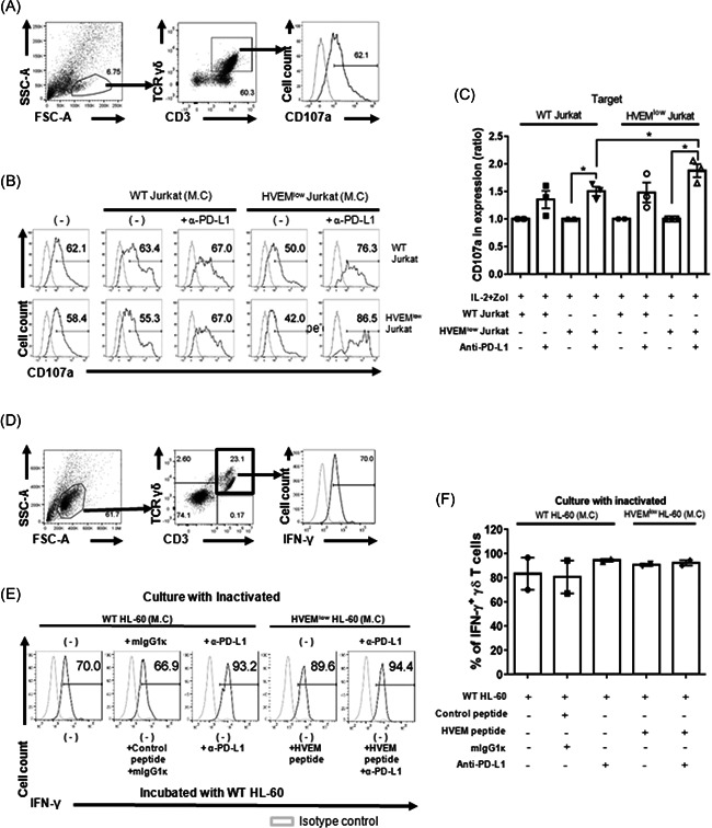
Figure 4.

Blockade of the BTLA/HVEM signaling does not affect the degranulation of in vitro expanded γδ T cells. (A) Representative histograms showing CD107a expression in the γδ T cells. Gray lines indicate the isotype control. (B) Cells were expanded in the indicated culture condition for 14 days. In the presence of anti‐human CD107a mAb, in vitro expanded γδ T cells with the indicated inactivated cells (on top) were incubated overnight with WT Jurkat (upper panel) or HVEMlow Jurkat (lower panel) cells. Representative data are shown out of three to four independent experiments using distinct donors. (C) The percentages of CD107a+ γδ T cells in the anti‐PD‐L1 treated group were normalized by dividing by that of the untreated group. Data were shown as mean ± SEM of three to four independent experiments using distinct donors. Paired t test was used (*p < .05). Each P value is .0190, .0345, and .0103 from left to right. (D) The gating strategy to identify intracellular IFN‐γ is depicted. (E) Expanded γδ T cells with the indicated inactivated cells (on top) were incubated overnight with WT Jurkat cells in the presence of BTLA/HVEM blocking peptides and anti‐PD‐L1 Ab or adequate controls for IFN‐γ intracellular staining. (F) The percentages of IFN‐γ+ γδ T cells were summarized. N = 2. BTLA, B‐ and T‐lymphocyte attenuator; HVEM, herpes virus entry mediator; IFN‐γ, interferon; mAB, monoclonal antibody; PD‐L1, programmed death‐ligand 1; WT, wild‐type
