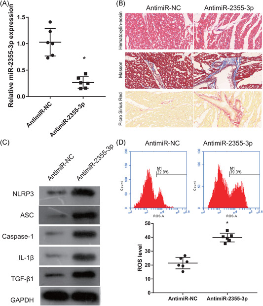
Figure 4.

MicroR‐2355‐3p reverses SOX2‐OT effects in rats with VA‐HF. (A) SOX2‐OT expression measured by qRT‐PCR 6 weeks after coinjection of si‐SOX2‐OT and anti‐miR‐2355‐3p. (B) Ventricular chambers stained with H&E, Masson trichrome, and Picro Sirius Red 6 weeks after coinjection of si‐SOX2‐OT and anti‐miR‐2355‐3p (200×). (C) Caspase‐1, NLRP3, ASC, IL‐1β, and TGF‐β1 expression measured by Western blot analysis 6 weeks after coinjection of si‐SOX2‐OT and anti‐miR‐2355‐3p. (D) Levels of ROS measured by flow cytometry 6 weeks after coinjection of si‐SOX2‐OT and anti‐miR‐2355‐3p. Anti‐miR‐NC group: coinjected with si‐SOX2‐OT and anti‐miR‐2355‐3p negative control; anti‐miR‐2355‐3p group: coinjected with si‐SOX2‐OT and anti‐miR‐2355‐3p. GAPDH, glyceraldehyde 3‐phosphate dehydrogenase; HF, heart failure; H&E, hematoxylin‐eosin; IL‐1β, interleukin‐1β; NLRP3, nucleotide‐binding oligomerization domain‐like receptor family pyrin domain containing 3; miR, microRNA; qRT‐PCR, quantitative reverse transcriptase‐polymerase chain reaction; ROS, reactive oxygen species; si, small interfering; SOX2‐OT, SOX2‐overlapping transcripts. *p < .05 (n = 6 per group)
