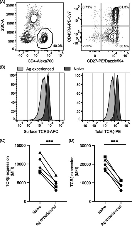
Figure 1.

Reduced TCR expression in antigen‐experienced primary human CD4+ T cells. Human PBLs from healthy donors were gated (A) to select CD4+, CD45RA+CD27+, naive and CD45RA−CD27− antigen‐experienced T cells and measure surface TCRβ and total TCRζ expression within these separate populations (B). Data in (A) and (B) are one representative experiment of the five biological replicates depicted in (C) and (D). (C,D) Aggregate MFI data for surface TCRβ (C) and total TCRζ (D) expression for n = 5 donors in one experiment, with each symbol representing an individual donor. MFI, mean fluorescence intensity; PBL, peripheral blood lymphocytes; TCR, T‐cell receptor. Paired Student's t test, ***p < .001
