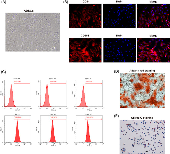
Figure 1.

Culture, isolation and identification of ADSCs. (A), cell morphologies under microscope after isolated cells being culture for respectively 24 h (left) and 14 days (right); (B), detection of biomarkers of ADSCs, CD44 (left) and CD105 (right) using immunofluorescent staining; (C), FCM measured the expressions of CD105, CD166, CD44, CD116, CD34, and CD45 in the 4th generation of ADSCs; (D), osteogenesis differentiation potential of ADSCs was verified using alizarin red staining; (E), adipogenic differentiation potential of ADSCs was verified using oil red O staining; ADSCs, adipose‐derived stem cells; FCM, flow cytometry
