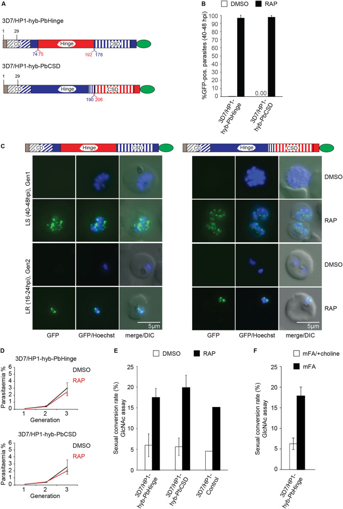
FIG 4.
Generation of DiCre-inducible PfHP1-PbHP1 hybrid mutants. (A) Diagrams showing the GFP-tagged PfHP1-PbHP1 hybrid proteins expressed in the 3D7/HP1-hyb-PbHinge and 3D7/HP1-hyb-PbCSD cell lines after RAP treatment. Brown and blue colors represent the remaining wild-type PfHP1 N terminus and the replacing PfHP1 protein sequences, respectively. Red colors identify the hinge domain and CSD derived from PbHP1. The CD and CSD are indicated by diagonal and vertical dashed stripes, respectively. Numbers in blue and red refer to amino acid positions within the PfHP1 and PbHP1 sequences, respectively. (B) Proportions of GFP-positive parasites observed 40 h after treatment with RAP or DMSO (control). Values represent the means from three independent biological replicates (error bars indicate SD). For each sample, >140 iRBCs were counted. (C) Representative live-cell fluorescence images showing the localization of GFP-tagged PfHP1-PbHP1 hybrid proteins in 3D7/HP1-hyb-PbHinge and 3D7/HP1-hyb-PbCSD parasites in late schizonts (LS) (40 to 48 hpi; generation 1 [Gen1], 40 h after RAP treatment) and in late-ring-stage progeny (LR) (16 to 24 hpi, generation 2). Nuclei were stained with Hoechst dye. DIC, differential interference contrast. Scale bar, 5 μm. (D) Growth curves of the DMSO- and RAP-treated 3D7/HP1-hyb-PbHinge and 3D7/HP1-hyb-PbCSD parasites over three consecutive generations. Values are the means from at least three independent replicate experiments (error bars represent SD). For each sample, >3,000 RBCs were counted. (E) Sexual conversion rates of the DMSO- and RAP-treated 3D7/HP1-hyb-PbHinge and 3D7/HP1-hyb-PbCSD mutants and the 3D7/HP1-Control line, assessed by inspection of Giemsa-stained blood smears of GlcNAc-treated cultures on day 6 of gametocytogenesis. Values represent the means from at least three independent replicate experiments (error bars represent SD). The values for the 3D7/HP1-Control line derive from a single experiment and are consistent with previously published data (69). For each sample, >3,000 RBCs were counted. (F) Sexual conversion rates of 3D7/HP1-hyb-PbHinge parasites cultured in minimal fatty acid medium (mFA) or mFA supplemented with 2 mM choline (mFA/+choline), assessed by inspection of Giemsa-stained blood smears of GlcNAc-treated cultures on day 6 of gametocytogenesis. Values represent the means from two independent replicate experiments (error bars represent SD). For each sample, >1,800 RBCs were counted.

