
Figure 1.
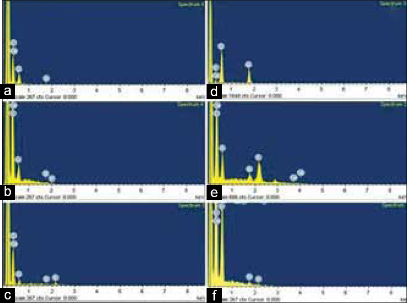
Energy-dispersive X-ray spectroscopy elemental spectrum (a) Energy-dispersive X-ray spectroscopy elemental spectrum of demineralized enamel sample (b) After nanohydroxyapatite infiltration in enamel sample (c) After nSiO2 infiltration in enamel sample (d) Spectrum of demineralized dentin sample (e) After nanohydroxyapatite infiltration in dentin sample (f) After nSiO2 infiltration in dentin sample