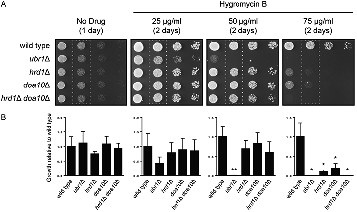
Figure 1. Loss of UBR1 sensitizes yeast to hygromycin B.

(A) Six-fold serial dilutions of yeast of the indicated genotypes were spotted onto agar plates containing rich medium (No Drug) or rich medium containing increasing concentrations of hygromycin B. Plates were imaged after 1-2 days (as indicated) of incubation at 30°C. (B) Growth in the second column of each plate (dashed rectangles) from three replicate experiments was quantified by densitometry. Data were analyzed by one-way ANOVA followed by Tukey post-hoc analysis (*, less than wild type; **, less than wild type, hrd1Δ, doa10Δ, and hrd1Δ doa10Δ; p < 0.05). Error bars represent standard error of the mean.
