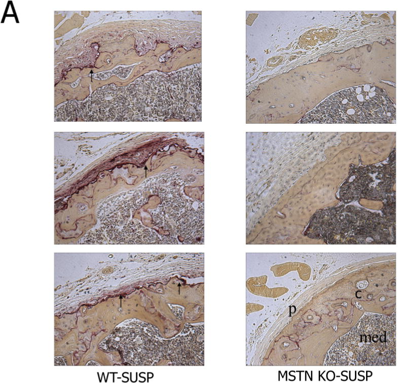
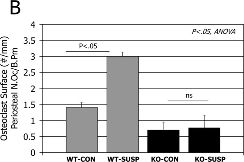
Figure 2.


A. Histological sections of the distal femur stained for tartrate resistant acid phosphatase in normal mice (WT, left column) and mice lacking myostatin (MSTN KO, right column) after 7 days hindlimb unloading (SUSP). Arrows point to numerous osteoclasts on the subperiosteal surface in wild-type mice but not in myostatin-deficient mice after unloading. C=cortical bone, p=periosteum, med=medullary cavity. B. Quantification of osteoclast number per bone surface on the periosteum of ground control (CON) and tail-suspended (SUSP) normal (WT) and myostatin-deficient (MSTN KO) mice.
