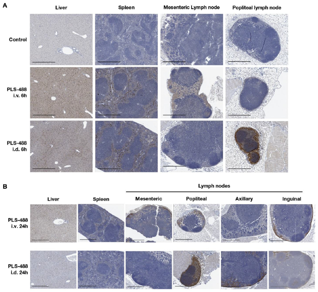
Figure 5.

(A) Displayed are the anti-Alexa488 immunohistochemistry images for liver, spleen, mesenteric lymph node and popliteal lymph node from mice 6 h after PBS vehicle control treatment and i.v. and i.d. injection of PLS-488. (B) Displayed are the anti-Alexa488 IHC images for liver, spleen, mesenteric lymph node, popliteal lymph node, axillary lymph node, and inguinal lymph node from mice 24 h after i.v. and i.d. PLS-488 treatment. The scale bars in the images are 500 um.
