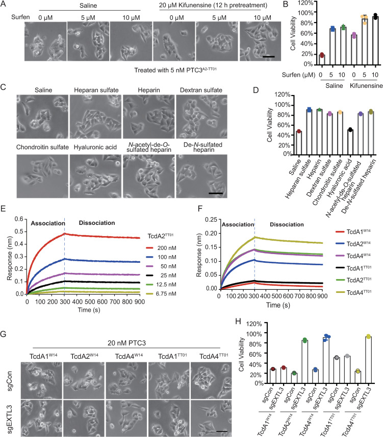
Fig 5. TcdA2TT01 binds with the sulfate group of sulfated glycosaminoglycans.
(A-B) HeLa-Cas9-sgCon cells pretreated with or without 20 μM Kifunensine for 12 h were exposed to 0, 5, 10 μM Surfen, respectively. Representative bright field micrographs (A) and cell viability (B) are shown. (C-D) HeLa-Cas9-sgCon cells were pre-incubated with 20 μM Kifunensine for 12 h and the indicated glycosaminoglycans (all at 1 mg/ml) for 1 h, then treated with 5 nM PTC3A2-TT01. Representative bright field micrographs (C) and the effects on cell viability (D) are shown. (E-F) BLI sensorgrams of TcAs interacting with immobilized biotin-Heparin. (E) BLI sensorgrams of TcdA2TT01 interacting with biotin-Heparin. TcA pentamer concentrations were 6.75–200 nM. (F) The biosensors loaded with biotin-Heparin were exposed to 50 nM of the indicated TcAs, followed by washing in HBS-EP buffer. The association and dissociation phases of each group are separated by a dashed line. The Kinetic parameters were listed in S5C Fig. (G-H) Representative bright field micrographs of HeLa-Cas9-sgCon cells treated with indicated Tc toxins (G). Cell viability was measured using CCK-8 assays and shown in (H).

