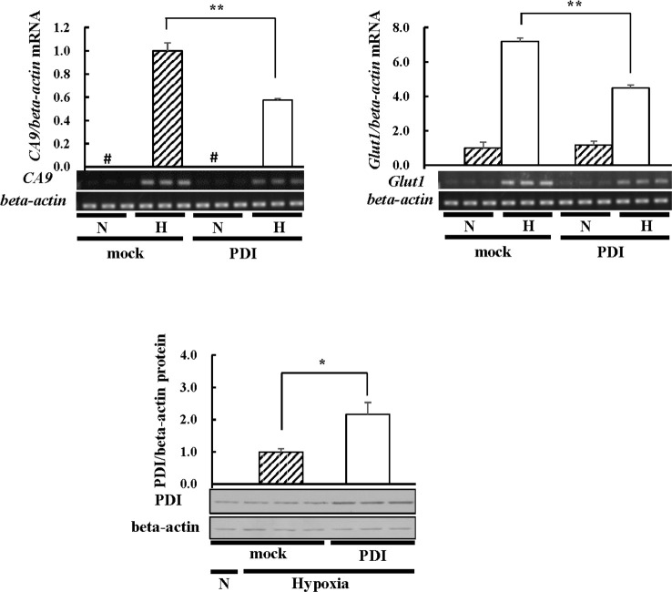
Fig 1. CA9 and Glut1 mRNA levels in PDI-overexpressing cells.
Hep3B cells overexpressing PDI were cultured for 6 h under hypoxic conditions. Total RNA was isolated from cells in three different plates, and the mRNA levels of CA9 or Glut1 were assessed by RT-PCR. The Glut1 mRNA levels of mock cells under normoxic conditions or the CA9 mRNA levels of mock cells under hypoxic conditions were set to 1.0. Proteins (3 μg) were separated by SDS-PAGE and immunoblotting was performed with an anti-PDI or -beta-actin antibody (1:1000 dilution). The PDI protein levels of mock cells under hypoxic conditions were set at 1.0. Values are expressed as the mean ± S.D. (error bars) of three different plates. N, Normoxia; H, Hypoxia. * p<0.05; ** p<0.01, significantly different from mock cells under hypoxic conditions. #, less than 0.05.

