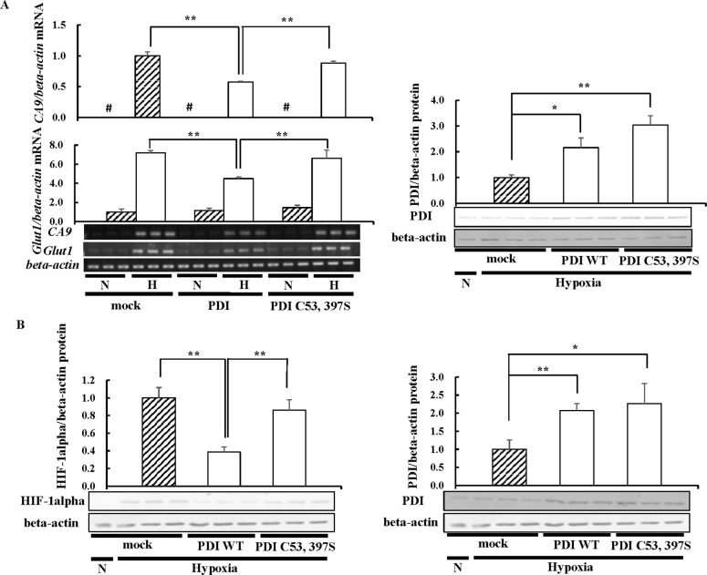
Fig 3. HIF-1alpha protein levels in catalytic inactive mutant PDI-overexpressing cells.
(A) Hep3B cells overexpressing PDI WT or PDI C53, 397S were cultured for 6 h under hypoxic conditions, and the mRNA levels of CA9 or Glut1 were assessed by RT-PCR. (B) Hep3B cells overexpressing PDI WT or PDI C53, 397S were cultured for 6 h under hypoxic conditions and then subjected to immunoblotting. The overexpression of PDI WT or PDI C53, 397S in Hep3B cells was assessed by Western blotting with an anti-PDI antibody (right panel) (A and B). The HIF-1alpha or PDI protein levels of mock cells under hypoxic conditions were set to 1.0. Values are expressed as the mean ± S.D. (error bars) of three different plates. N, Normoxia; H, Hypoxia. * p<0.05; ** p<0.01, significantly different from mock cells under hypoxic conditions. #, less than 0.05.

