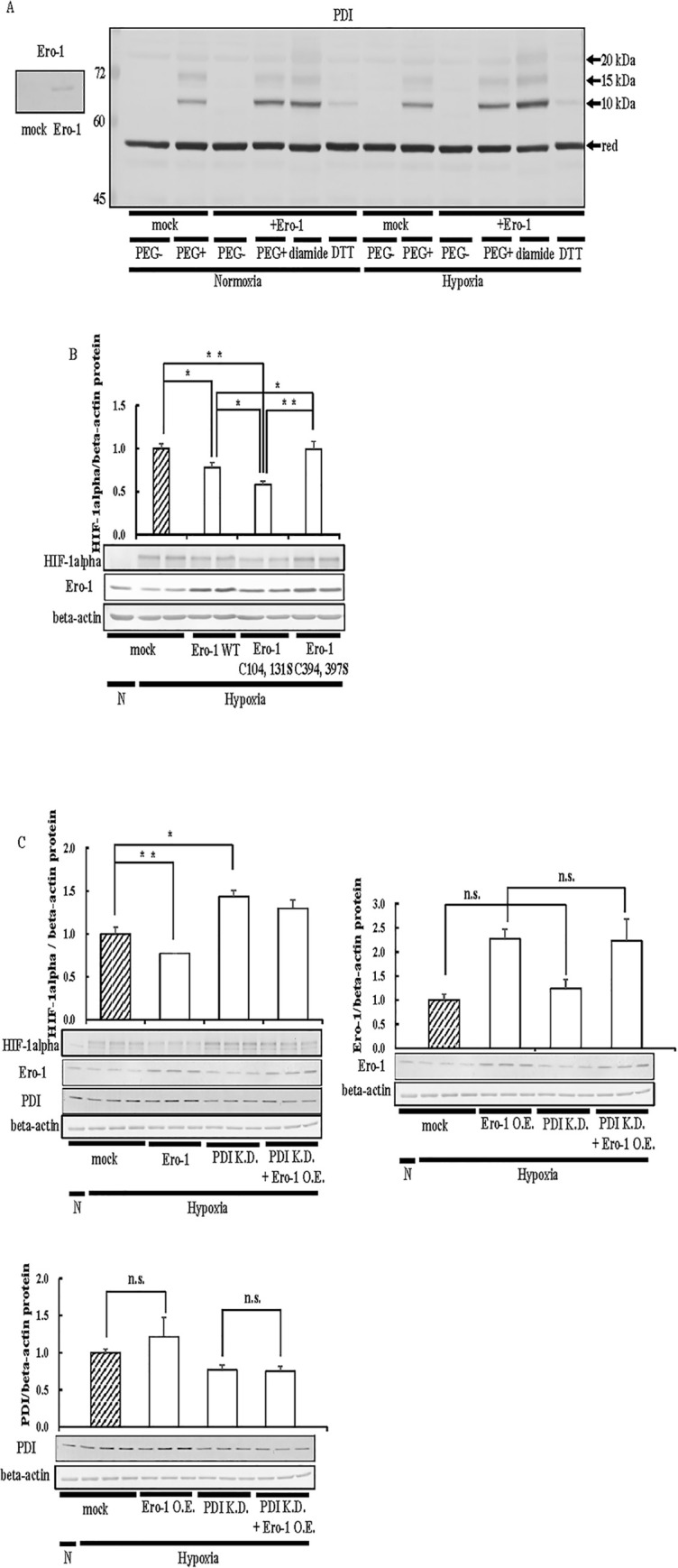
Fig 4. Effects of Ero-1 on HIF-1alpha expression.

(A) Hep3B cells overexpressing Ero-1 WT were cultured for 6 h under hypoxic conditions. Cells were harvested with PBS, and proteins were incubated in the absence or presence of 5 mM diamide or 10 mM DTT. Precipitated proteins were incubated with 20 mM NEM. After the removal of NEM, proteins were incubated with 1 mM DTT. Precipitated proteins were incubated with 1 mM 5kDa PEG-maleimide. An up-shift in molecular weight by the binding of PEG-maleimide was detected by immunoblotting with the anti-PDI antibody. To evaluation of the overexpression of Ero-1 (left panel), proteins (3 μg) were separated by SDS-PAGE, and immunoblotting was performed with the anti-Ero-1 antibody (1:1000 dilution). red, Reduced form. (B) Hep3B cells overexpressing Ero-1 WT or Ero-1 C104, 131S, and Ero-1 C394, 397S were cultured for 6 h under hypoxic conditions, and immunoblotting was performed. (C) Ero-1/pcDNA 3.1 (+) and si-PDI were transfected into Hep3B cells, cells were cultured for 6 h under hypoxic conditions, and immunoblotting was performed. The overexpression of Ero-1 or PDI was evaluated by immunoblotting with an anti-Ero-1 or PDI antibody (1:1000 dilution). The HIF-1alpha, Ero-1, or PDI protein levels of mock cells under hypoxic conditions were set to 1.0. Values are expressed as the mean ± S.D. (error bars) of three different plates. N, Normoxia; H, Hypoxia; n. s., not significant. * p<0.05; ** p<0.01, significantly different from mock cells under hypoxic conditions.
