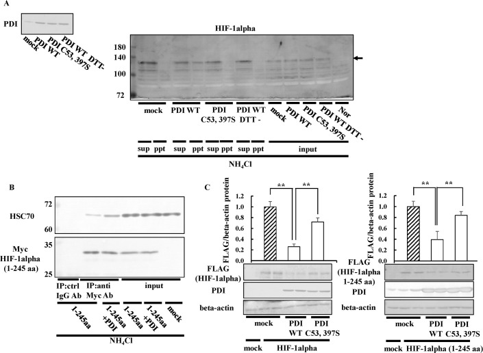
Fig 9. Effects of PDI on HIF-1alpha (1–245 aa) expression.
(A) Hep3B cells overexpressing PDI WT or PDI C53, 397S were cultured under hypoxic conditions for 8 h in the presence of NH4Cl. The biotin-switch method was performed, and HIF-1alpha was detected by immunoblotting with the anti-HIF-1alpha antibody. (B) Hep3B cells overexpressing HIF-1alpha 1–245 aa or PDI were cultured for 8 h in the presence of NH4Cl. Cell extracts were subjected to immunoprecipitation using the anti-Myc antibody (1:1000 dilution). Precipitated proteins (15 or 3 μg) were separated by SDS-PAGE, and immunoblotting was performed with the anti-Myc or -HSC70 antibody. (C) HIF-1alpha full length or 1–245 aa/3×FLAG-pcDNA4 vector, or PDI WT, or PDI C53, 397S / pcDNA 3.1 (+) was transfected into Hep3B cells, and immunoblotting was performed with the anti-FLAG, -PDI, or-beta-actin antibody. The HIF-1alpha protein levels of mock cells under normoxic conditions were set to 1.0. Values are expressed as the mean ± S.D. (error bars) of three different plates. ctrl, control. ** p<0.01, significantly different from mock cells.

