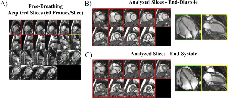
Fig 1. Imaging processing for free-breathing, real-time acquisitions.
(A) All slices (10–12 SAX and 6 each of 2 and 4-chamber LAX) are viewed simultaneously for selection of those for analysis. All SAX slices with myocardium and a single 2- and 4-chamber LAX are selected for analysis. A full cardiac cycle for each selected slice is extracted, from which end-diastolic (B) and end-systolic (C) images are identified and endocardial (red) and epicardial (green) borders are traced.

